human anatomy humor
A living 28 week old human fetus photographed in the womb in 1965 by. 9 Pics about A living 28 week old human fetus photographed in the womb in 1965 by : A living 28 week old human fetus photographed in the womb in 1965 by, Cat Dissection Study Tools | Vet medicine, Cat anatomy, Anatomy and also Student Study Guides - Skull Anatomy.
A Living 28 Week Old Human Fetus Photographed In The Womb In 1965 By
 www.pinterest.com
fetus womb week lennart human medical nilsson 1965 embryo illustration comfort zone living photographed
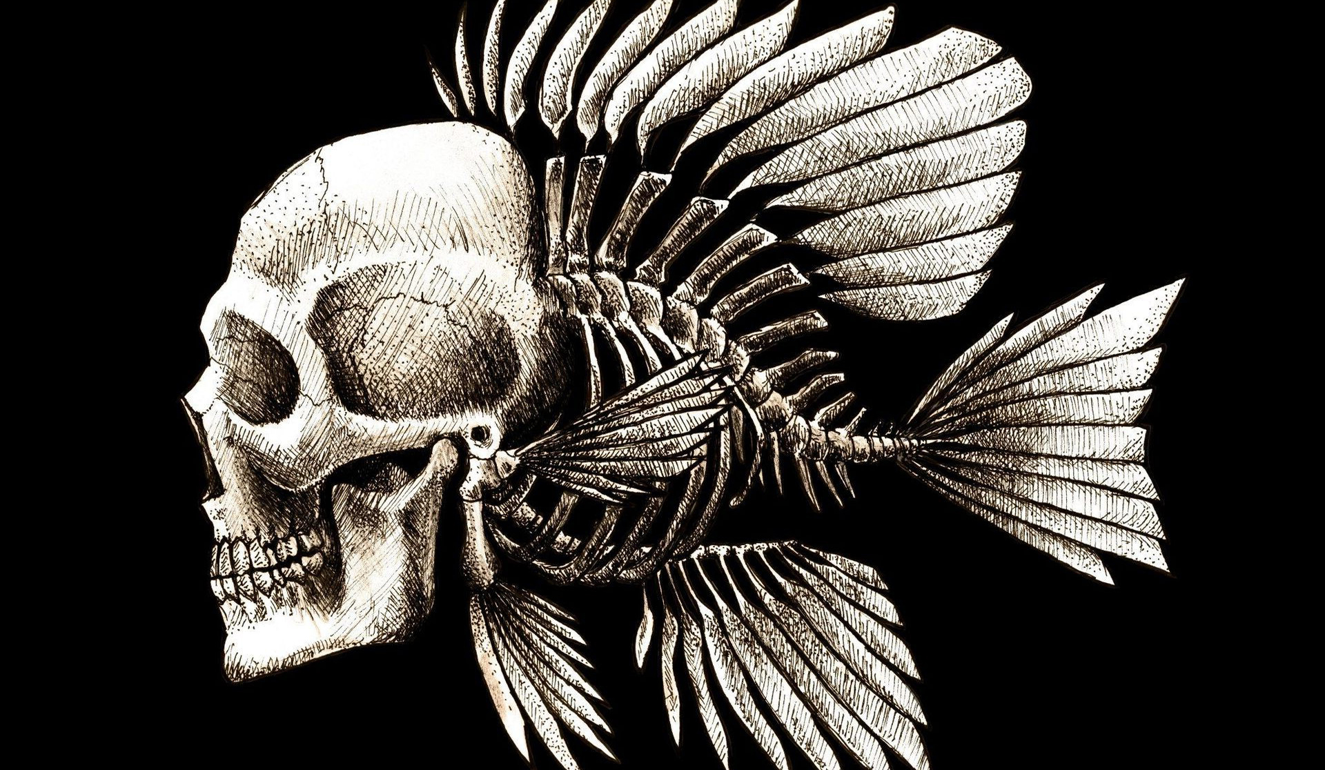
Skulls Humor Fish Artwork Charles Darwin Bones Seaman Wallpapers HD
 wallup.net
darwin charles fish wallpapers bones skeleton seaman disturbing artwork skull human skulls desktop backgrounds humor skelton abstract background body mobile
Cute Illustrations Help Kids Identify Different Parts Of The Human
 designtaxi.com
parts anatomy human cute body cartoon illustrations identify different help system skeletal systems designtaxi illustration science heart bodies biology physiology
Elbow - Radiography (Anterior-posterior View) : Lateral Epicondyle
 www.pinterest.com
elbow anatomy ap radiography upper lateral joint radiology ray anterior radiograph posterior limb mri humerus olecranon imaios coronoid trochlea medical
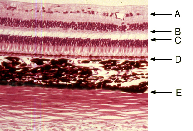
Eye | Histology
 histology.medicine.umich.edu
histology eye questions umich medicine edu
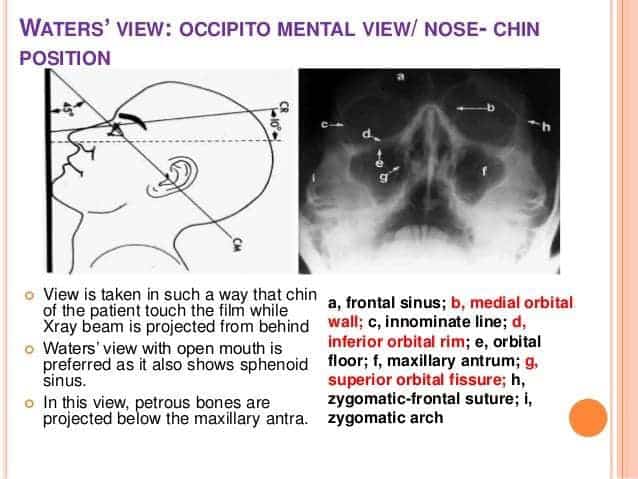
Student Study Guides - Skull Anatomy
 theradiologictechnologist.com
positioning radiology bones
Instant Anatomy - Abdomen - Surface - Transpyloric Plane | Nursing
 www.pinterest.com
anatomy surface transpyloric plane abdomen instant planes body abdominal vertebral aorta instantanatomy diagrams arteries
MRI Brain Ambiens Cistern Anatomy | Radiology Anatomy Images | Mri
 www.pinterest.com
mri cistern anatomy brain ambiens radiology coronal ct imaging sectional cross neurology visit
Cat Dissection Study Tools | Vet Medicine, Cat Anatomy, Anatomy
 www.pinterest.com
cat anatomy dissection system excretory veins human circulatory body animal study tools loved medicine arteries vet veterinary
Anatomy surface transpyloric plane abdomen instant planes body abdominal vertebral aorta instantanatomy diagrams arteries. Cute illustrations help kids identify different parts of the human. Cat dissection study tools