hip bone anatomy axial
PELVIS AND UPPER FEMORA | Radiology Key. 9 Pics about PELVIS AND UPPER FEMORA | Radiology Key : PELVIS AND UPPER FEMORA | Radiology Key, Femur bone anatomy: Proximal, distal and shaft | Kenhub and also Hip and Pelvis | Musculoskeletal Key.
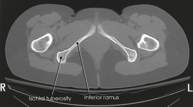
PELVIS AND UPPER FEMORA | Radiology Key
 radiologykey.com
pelvis ct anatomy axial ramus ischial inferior tuberosity upper femora radiology sectional imaging petersen kelley modified fig cm radiologykey
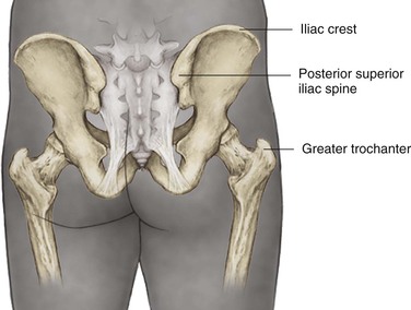
Hip And Pelvis | Musculoskeletal Key
 musculoskeletalkey.com
hip posterior iliac superior spine pelvis crest landmarks bony trochanter greater include around figure musculoskeletalkey
Stanford MSK MRI Atlas (c) 2020
shoulder mri anatomy atlas xrayhead pulley biceps msk stanford cuff rotator tear lecture labrum ankle
Female Pelvis (Anatomy) - Study Guide | Kenhub
:fill(FFFFFF,true):format(jpeg)/images/container/female-pelvis/Female_Pelvis_1.png) www.kenhub.com
www.kenhub.com
pelvis female kenhub anatomy
Normal Lumbar Spine MRI: 3 T | Image | Radiopaedia.org
 radiopaedia.org
lumbar spine mri normal axial t2 radiopaedia nerve version
Thoracic, Lumbar , Sacrum & Coccyx Vertebrae
 www.slideshare.net
sacrum coccyx lumbar vertebrae thoracic
Femur Bone Anatomy: Proximal, Distal And Shaft | Kenhub
femur kenhub proximal coxae articulatio distal
Pin On Nursing School
 www.pinterest.com
humerus bone labeled anatomy labeling worksheets human google nursing bones studying example type biology skull gross skeleton
Acute L1 Burst Compression Fracture | Radiology Case | Radiopaedia.org
 www.pinterest.com
fracture radiopaedia lumbar medicine vertebrae radiograph fractures
Shoulder mri anatomy atlas xrayhead pulley biceps msk stanford cuff rotator tear lecture labrum ankle. Hip posterior iliac superior spine pelvis crest landmarks bony trochanter greater include around figure musculoskeletalkey. Stanford msk mri atlas (c) 2020